Fifteen amazing body shots captured using a scanning electron microscope,
showing incredible details of 1 to 5 nm (nanometers) in size.
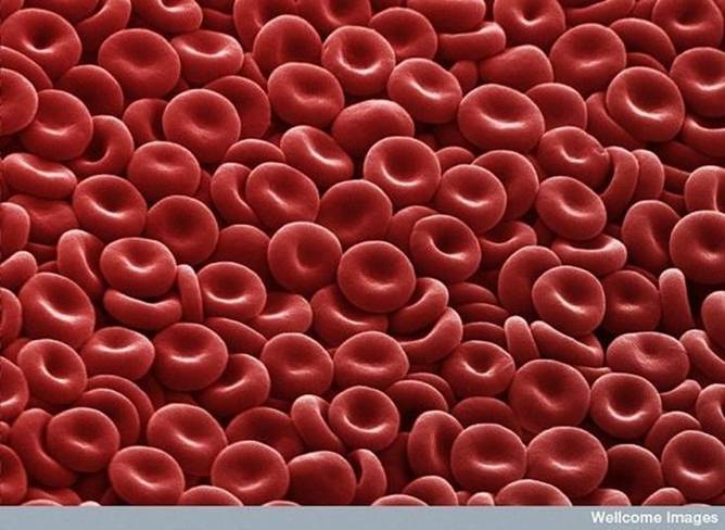
1. Red Blood Cells
They look like little cinnamon candies here, but they're actually the most common type of blood cell in the human body - red blood cells (RBCs). These biconcave-shaped cells have the tall task of carrying oxygen to our entire body; in women there are about 4 to 5 million RBCs per micro liter (cubic millimeter) of blood and about 5 to 6 million in men. People who live at higher altitudes have even more RBCs because of the low oxygen levels in their environment.
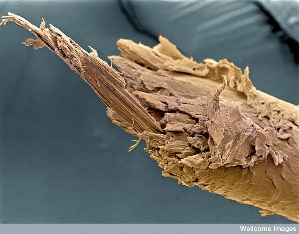
2. Split End of Human Hair
Regular trimmings to your hair and good conditioner should help to prevent this unsightly picture of a split end of a human hair.
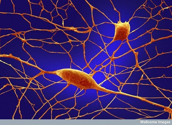
3. Purkinje Neurons
Of the 100 billion neurons in your brain. Purkinje (pronounced purr-kin-jee) neurons are some of the largest. Among other things, these cells are the masters of motor coordination in the cerebellar cortex. Toxic exposure such as alcohol and lithium, autoimmune diseases, genetic mutations including autism and neurodegenerative diseases can negatively affect human Purkinje cells.
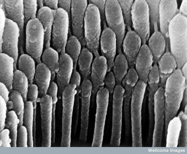
4. Hair Cell in the Ear
Here's what it looks like to see a close-up of human hair cell stereo cilia inside the ear. These detect mechanical movement in response to sound vibrations.
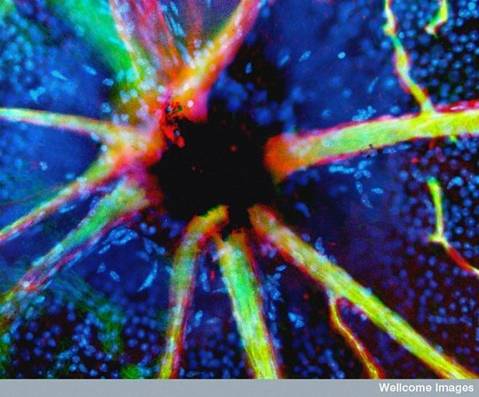
5. Blood Vessels Emerging from the Optic Nerve
In this image, stained retinal blood vessels are shown to emerge from the black-colored optic disc. The optic disc is a blind spot because no light receptor cells are present in this area of the retina where the optic nerve and retinal blood vessels leave the back of the eye.
6. Tongue with Taste Bud
This colour-enhanced image depicts a taste bud on the tongue. The human tongue has about 10,000 taste buds that are involved with detecting salty, sour, bitter, sweet and savory taste perceptions. Thai people have very few -- most killed by eating spicy food.
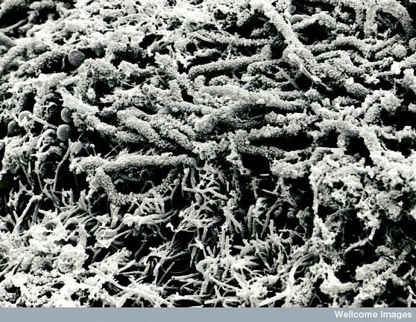
7. Tooth Plaque
Brush your teeth often because this is what the surface of a tooth with a form of corn-on-the-cob plaque looks like.
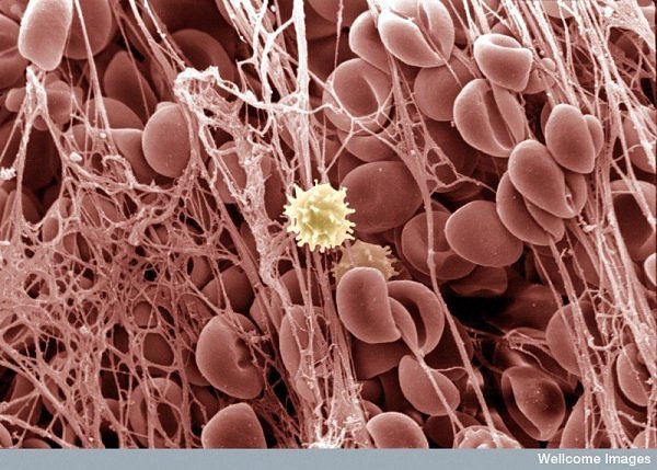
8. Blood Clot
Remember that picture of the nice, uniform shapes of red blood cells you just looked at? Well, here's what it looks like when those same cells get caught up in the sticky web of a blood clot. The cell in the middle is a white blood cell.
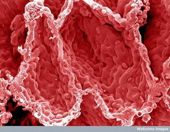
9. Alveoli in the Lung
This is what a colour-enhanced image of the inner surface of your lung looks like. The hollow cavities are alveoli; this is where gas exchange occurs with the blood.
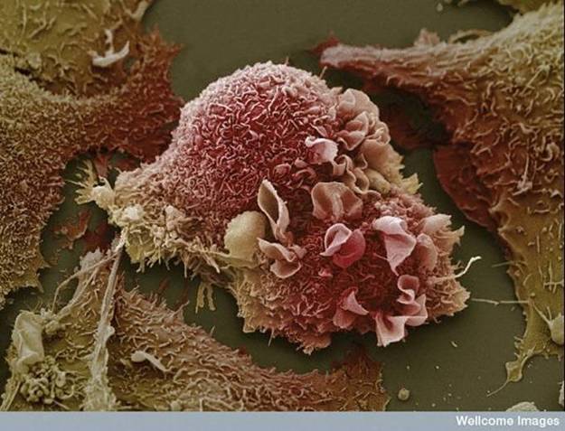
10. Lung Cancer Cells
This image of warped lung cancer cells is in stark contrast to the healthy lung in the previous picture.
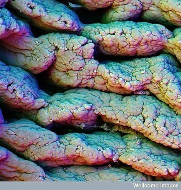
11. Villi of Small Intestine
Villi in the small intestine increase the surface area of the gut, which helps in the absorption of food. Look closely and you will see some food stuck in one of the crevices.
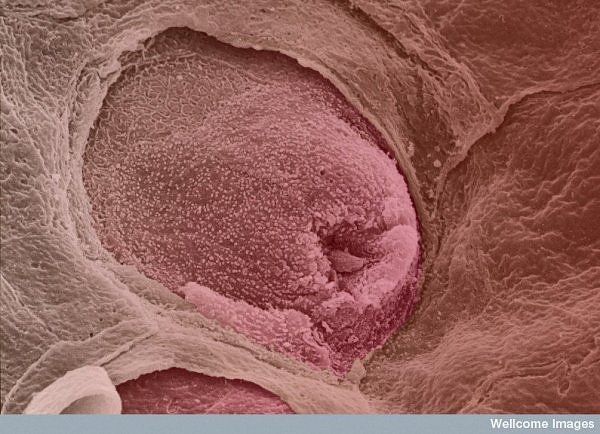
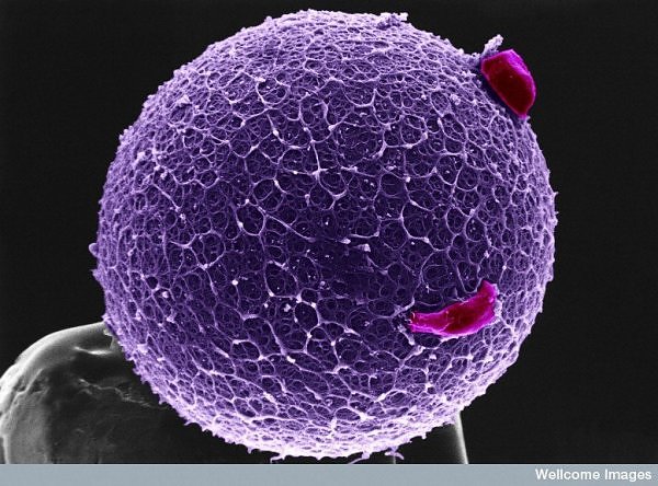
12. Human Egg with Coronal Cells
This image is of a purple, colour-enhanced human egg sitting on a pin. The egg is coated with the zona pellicuda, a glycoprotein that protects the egg but also helps to trap and bind sperm. Two coronal cells are attached to the zona pellicuda.
Images © Wellcome Library, London
13. Sperm on the Surface of a Human Egg
Here's a close-up of a number of sperm trying to fertilize an egg.
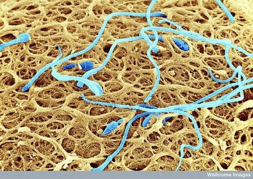
14. Human Embryo and Sperm
It looks like the world at war, but it is actually five days after the fertilisation of an egg, with some remaining sperm cells still sticking around. This fluorescent image was captured using a confocal microscope. The embryo and sperm cell nuclei are stained purple while sperm tails are green. The blue areas are gap junctions, which form connections between the cells.
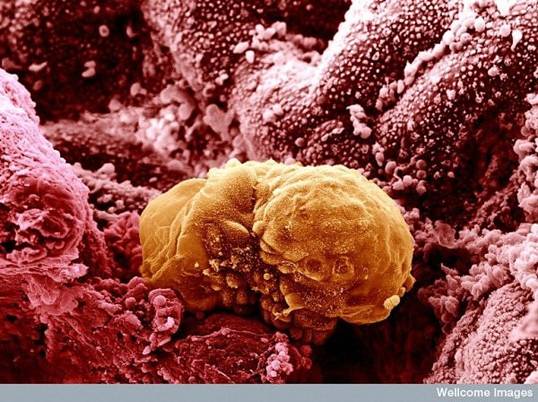
15. Colored Image of a 6 day old Human Embryo Implanting itself onto the wall of the womb.